Nanoscopic XAFS-CT (X-ray Absorption Fine Structures Computed Tomography): three-dimensional visualization of chemical states with a high spatial resolution
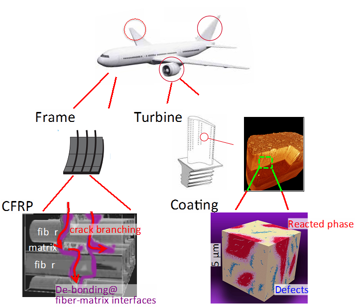
The macroscopic performance of advanced materials is often associated with various reactions which progress heterogeneously. Thus, material properties are generally determined not by their averaged characteristics but by specific features in heterogeneity (or ‘trigger sites’) of phases, chemical states, etc., where the key reactions dictate macroscopic properties. We have investigated trigger sites in structural materials, batteries, etc. using X-ray microscopy.
Some of ongoing projects include: (a) the in situ observation of crack formation in multi-scales of FOV (from a few μm to a few tens of mm) in CFRP and ceramic coatings using X-CT, (b) chemical state mapping using X-ray absorption in multi-scale FOV (from a few μm to a few tens of mm) in oxide, ceramic coatings, and CFRP, (c) application of informatics and/or applied mathematics to the handling of multi-dimensional data such as chemical states and/or microstructures as a function of space (3D), energy, and time (Fig. 1).
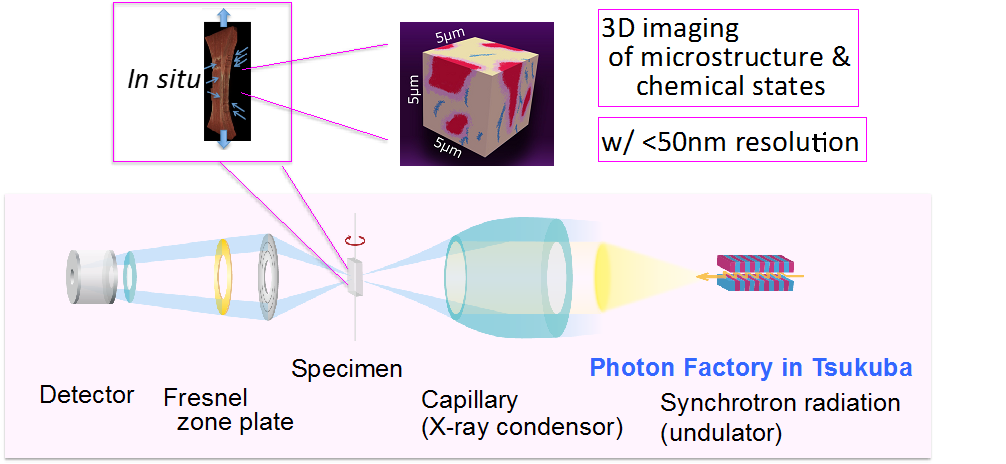
New X-ray microscopy equipment (XAFS-CT) will be installed in the beam line of AR-NW2A, in PF-AR, KEK in FY2016; with this equipment, an X-ray beam will be focused onto a specimen through a capillary condenser, and a full-field transmission image will be obtained using a zone-plate. By scanning the X-ray energy, we will be able to obtain 3D X-CT images with X-ray absorption near-edge structure (XANES) spectroscopy (Fig. 2).